First Electronic Microscopic image of Covid- 19 in India
Pune: The images of the Covid- 19 have been taken by a team of ICMR-NIV scientists in Pune.
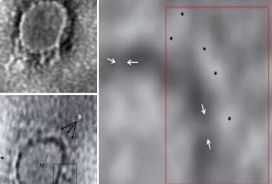
The first images from India of what the Covid- 19 looks like are out. The images have been captured using a transmission electron microscope and have been published in the Indian Journal of Medical Research.
The images of Sars-Cov-2, the virus that causes COVID-19, are from the throat swab of the first laboratory confirmed case in India reported on January 30. The woman, among three students studying medicine in Wuhan in China, was diagnosed with COVID-19 after returning to India.
The gene sequencing of the samples from Kerala done at the National Institute of Virology (NIV) in Pune found that the virus was a 99.98 per cent match with the virus in Wuhan. The image revealed the presence of stalk-like projections ending in round structures typical of a Covid- 19 particle.
The article titled “Transmission electron microscopy imaging of SARS-CoV-2” has been authored by the ICMR-NIV National Influenza Centre Team. The authors include Atanu Basu, deputy director and head of electron microscopy and pathology at NIV Pune.
According to the study, the description of a novel human Corona virus, initially referred to as the Wuhan Corona virus (CoV), is currently designated as severe acute respiratory syndrome (SARS)-CoV-2 as per the latest International Committee on Taxonomy of Viruses (ICTV) classification. It is probably the most recent human pneumonia virus with high outbreak potential.